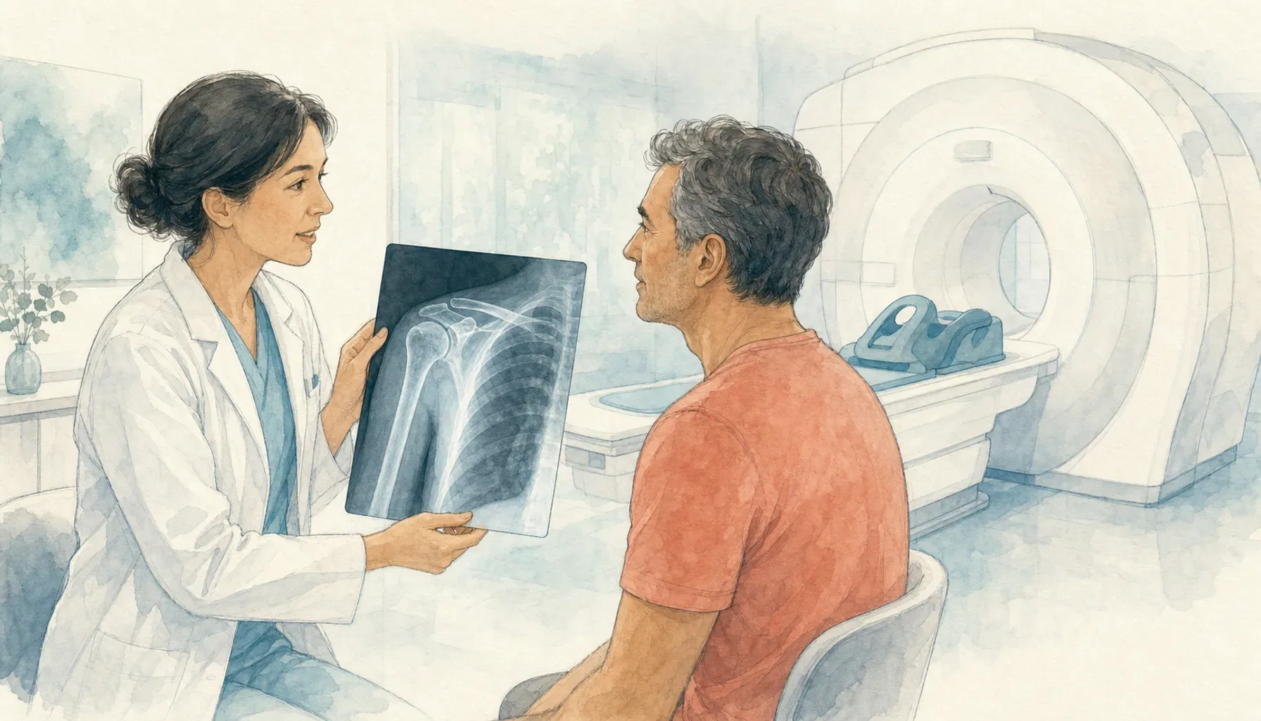
For many people with shoulder pain, X-rays are the first imaging study, while MRI is reserved for a specific soft-tissue or bone-marrow question after the history and examination. X-rays are well suited to fractures, alignment, arthritis, and other bone changes. MRI can show the rotator cuff, labrum, muscles, and deeper soft tissues.
The right choice depends on whether pain followed trauma, how long it has lasted, which movements are limited, and what the clinician suspects. An expensive scan is not automatically a better starting point.
X-ray and MRI at a glance
| Feature | Shoulder X-ray | Shoulder MRI |
|---|---|---|
| Shows best | Bone, joint alignment, arthritis, many fractures, calcification | Tendons, muscles, labrum, cartilage, marrow, fluid |
| Radiation | Uses a small dose of ionizing radiation | Does not use ionizing radiation |
| Typical role | Often the initial study for acute or chronic pain | Selected after examination and often after X-rays |
| Time and access | Usually quick and widely available | Longer exam with a confined scanner and safety screening |
| Main limitation | Does not directly show rotator cuff or labral tears | Can show findings that are unrelated to the current pain |
When an X-ray is often useful
The American College of Radiology rates shoulder radiography as usually appropriate for the initial imaging of chronic shoulder pain. X-rays can reveal osteoarthritis, calcific deposits, changes at the acromioclavicular joint, many fractures, bone lesions, and whether the joint is aligned.
After acute trauma, radiographs are also the usual first study because fractures and dislocations need to be identified promptly. If the X-rays are normal or unclear but the examination still suggests an occult fracture or soft-tissue injury, another test may follow.
X-rays cannot directly display tendons or the labrum. A normal X-ray therefore does not prove that every shoulder structure is normal. The comparison of shoulder pain and arthritis explains why bone and joint findings need to match the symptom pattern.
When MRI may answer the next question
MRI uses a strong magnetic field and radiofrequency energy to create detailed images. It can help evaluate the rotator cuff, biceps tendon, labrum, cartilage, marrow, muscles, and fluid. The ACR notes that MRI without intravenous contrast, ultrasound, or MR arthrography may be appropriate after normal or inconclusive radiographs when a particular soft-tissue condition is suspected.
The exact MRI protocol matters. A standard MRI and an MR arthrogram are not interchangeable. An arthrogram involves placing contrast into the joint and may be chosen for selected labral or instability questions. Intravenous contrast is usually unnecessary for routine rotator cuff evaluation, although infection, tumor, prior surgery, and other special situations can change the plan.
AAOS explains that an X-ray does not show the soft-tissue labrum and that MRI displays soft tissue better. For suspected rotator cuff disease, both MRI and diagnostic ultrasound can be useful adjuncts to the clinical examination. Availability, operator skill, prior surgery, implanted devices, and the exact clinical question affect the choice.
Imaging does not replace the examination
Shoulder abnormalities can appear on imaging in people who have little or no pain. A scan finding must be compared with the pain location, active and passive motion, strength, stability, tenderness, and neck or nerve signs. Treating an incidental image finding can send care in the wrong direction.
Tingling, numbness, reflex changes, or pain affected by neck movement may lead the clinician to evaluate the cervical spine and nerves. A shoulder MRI does not evaluate the whole nerve pathway. Read shoulder pain versus cervical radiculopathy and shoulder pain with hand tingling before assuming the scan should focus on the shoulder.
Questions to ask before a scan
Ask what diagnosis the test is meant to confirm or exclude, whether the result would change treatment, and whether another study could answer the question. Tell the imaging team about pregnancy, metal fragments, implanted electronic or medical devices, kidney disease, previous contrast reactions, and severe claustrophobia. MRI safety screening is essential.
Also ask how the results will be reviewed with you. The report is one part of the decision. Your clinician should connect the findings to symptoms and function. Persistent pain after several weeks warrants assessment, as discussed in shoulder pain at four weeks.
Care while you wait for evaluation
Avoid a movement that sharply increases pain and note what the shoulder can and cannot do. Do not begin forceful stretching or heavy strengthening based on an online diagnosis. New weakness, deformity, fever, redness, major swelling, or pain after significant trauma needs timely care.
If a clinician has approved hot or cold therapy for a musculoskeletal condition, a Shoulder Compression Wrap can hold a gel pack for short-term comfort. It cannot determine whether you need imaging or treat a fracture, tendon tear, infection, nerve problem, or tumor. Avoid temperature therapy when sensation or circulation is impaired unless a clinician gives specific guidance.
Call 911 for shoulder or arm pain with chest pressure, shortness of breath, sweating, nausea, or lightheadedness. Imaging decisions can wait until emergency causes have been addressed.
Common questions
Is an X-ray or MRI better for shoulder pain?
Each answers a different question. X-rays show bones, alignment, arthritis, and many fractures. MRI shows tendons, muscles, the labrum, bone marrow, and other soft tissues. The examination and suspected condition determine which is appropriate.
Can an X-ray show a rotator cuff tear?
A plain X-ray does not directly show the rotator cuff tendons. It may show arthritis, a bone spur, or other bone findings. Ultrasound or MRI may be selected when the examination suggests a rotator cuff tear.
Do you need an MRI for shoulder pain that will not go away?
Persistent pain deserves a clinical evaluation, but duration alone does not make MRI necessary. A clinician may start with an examination and X-rays, then choose MRI, ultrasound, CT, nerve testing, or no additional imaging according to the findings.
This article provides general education and cannot determine which imaging test is appropriate for you. Read our medical disclaimer. A licensed clinician and radiology team can select and interpret imaging in the context of your examination.